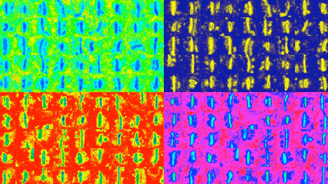
Electron
micrograph of stained somatosensory cortex synapses that were identified
using a
machine-learning algorithm. Image credit: Saket Navlakha and Alison L. Barth.
(December 29, 2015) High-throughput,
Machine-Learning Tool Could Help Researchers Better Understand Synaptic
Activity in Learning and Disease.
Carnegie Mellon University researchers have developed a new
approach to broadly survey learning-related changes in synapse properties.
In a study published in the Journal of Neuroscience and
featured on the journal’s cover, the researchers used machine-learning
algorithms to analyze thousands of images from the cerebral cortex. This
allowed them to identify synapses from an entire cortical region, revealing
unanticipated information about how synaptic properties change during
development and learning. The study is one of the largest electron microscopy
studies ever carried out, evaluating more subjects and more images than prior
researchers have attempted.
As the brain learns and responds to sensory stimuli, its
neurons make connections with one another. These connections, called synapses,
facilitate neuronal communication, and their anatomic and electrophysiological
properties contain information vital to understanding how the brain behaves in
health and disease. Researchers use different techniques, including electron
microscopy, to identify and analyze synapse properties. While electron
microscopy can be a useful tool for reconstructing neural circuits, it is also
data and labor intensive. As a result, researchers have only been able to use
it to study small, targeted areas of the brain until now.
Studying a large section of the brain using traditional
electron microscopy techniques would result in terabytes of unwieldy data,
given that the brain has billions of neurons, each with hundreds to thousands
of synaptic connections. The new technique developed at Carnegie Mellon
simplifies this problem by combining a specialized staining process with
machine learning.

